Pilates Therapy and Bodyworks

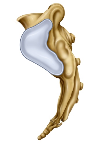
The auricular surface is located on the ilium and articulates with the same ear-shaped space on the lateral sides of the sacrum. The sacral surface comprises hyaline (the weakest type of collagen) cartilage, and the iliac surface is fibrocartilage (the most robust type of collagen). This connection makes up the sacroiliac joints. This joint is the least mobile synovial joint in the body. Unlike the pectoral girdle, made up to allow great ranges of movement, the pelvic girdle is mostly immobile. It is a transitional area from the spine to the pelvis and provides stability from the load of the trunk and weight transfer.

The joint surfaces are relatively flat in early life, allowing a small gliding motion in all directions. Reported movements are axial rotation of 1.5 degrees, lateral bending of 0.8 degrees, and flexion/extension, the largest at around 3 degrees. Women tend to have more mobility at the joints than men. As we age, the surface changes, becoming less flat with less glide. The main movement is anterior/posterior, known as nutation and counternutation. Nutation is when the sacrum moves anterior and inferior as the coccyx moves posteriorly relative to the ilium. Counternutation is where the opposite occurs.
The average movement at the joint is around 3 mm.
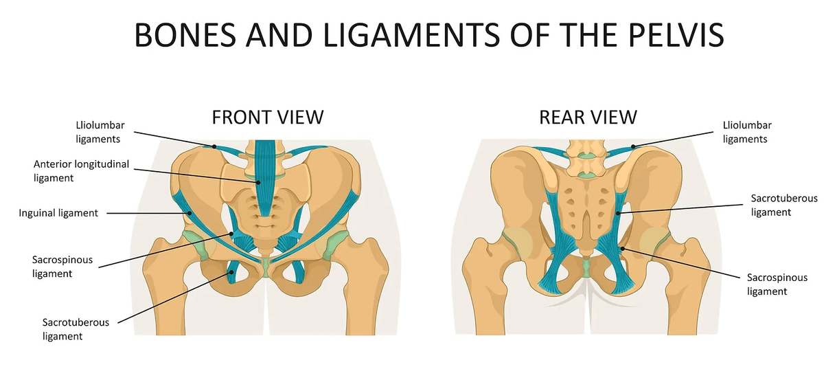
The interosseous sacroiliac ligaments (also referred to as the axial ligaments) are the significant connection and prevent nutation of the sacrum. The posterior sacroiliac ligaments resist counternutation being situated more superiorly. The sacrotuberous ligament blends into the inferior posterior sacroiliac ligaments to resist nutation. The anterior sacroiliac ligament weakly resists rotation and nutation. The sacrospinous ligaments resist rotation and nutation.

The pelvic floor connection. Engage your pelvic floor muscles. Perhaps you notice the tension on the coccyx? Imagine the tensioning as a slight pull forward of the coccyx, at the other end, the top of the sacrum tilting back; think of the tissues in these areas tensioning to resist these movements. The coccygeus muscle sits directly over the sacrospinous ligament and is often considered a degenerated part of the muscle.

