So many interesting things to look at when covering the anatomy of the body. Our fun fact for today is Cornu (a), something in the body that is horn-shaped, like, for instance, the horn-shaped projections on the hyoid bone.
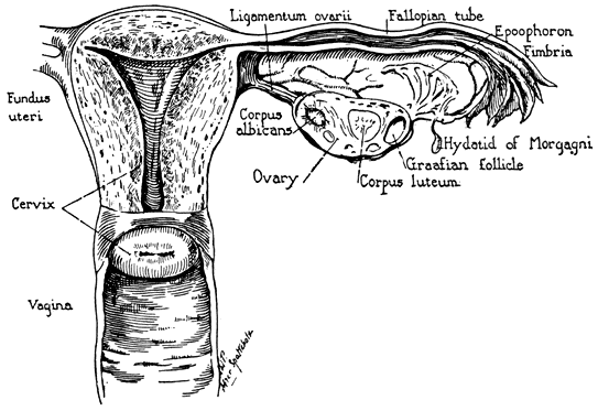
* Uterine Cornu – these projections meet with the fallopian tubes and the ovary at the top of the uterus.
(See clinical anatomy of the uterus, fallopian tubes, and ovaries. Sokol, E, Glob. libr. women’s med. (ISSN: 1756-2228) 2011; DOI 10.3843/GLOWM.100001) for a nice description of the female reproductive system and some great images.
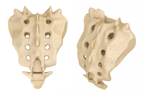
* Cornu coccygeum – these horn-shaped prominences at the top of the coccyx meet with the downward horn-shaped prominences of the sacrum. The 5th sacral nerves and a pair of coccygeal nerves pass through here along with filum terminale. This fibrous band extends from the lower spine from the conus medullaris to the periosteum of the coccyx. This band helps to stabilise and buffer the distal spinal cord as it tapers to become the horsetail like collection of nerve roots (cauda equina) from caudal traction and abnormal movements.
* Cornu anterius and posterius medullae spinalis. The grey matter of the spinal cord, the ventral/anterior division containing the alpha motor neurons, innervate the skeletal muscle, which causes movement. In addition, the dorsal/posterior horn contains neurons that receive somatosensory information. Finally, we have the intermediate or lateral horn, which contains neurons that innervate the visceral and pelvic organs.
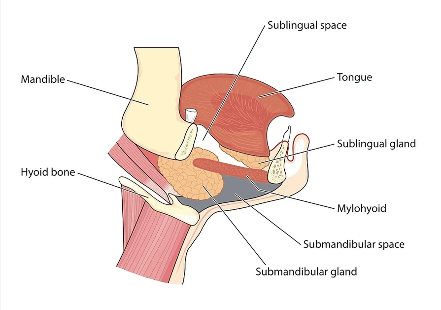
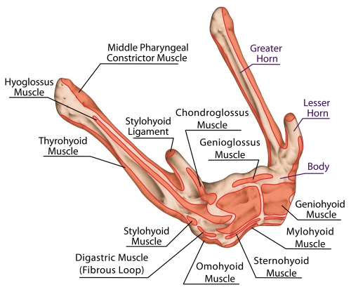
* Cornu of the Hyoid. U shape of the hyoid bone is made up of the body at the base, the longer and larger horns, the greater cornua and a pair of smaller horns, the lesser cornua at the junction of the greater horns and body. The hyoglossus muscle takes up the whole length of the greater horn; the middle pharyngeal constrictor also attaches to the greater horn. The stylohyoid ligament inserts onto the lesser Cornu of the hyoid bone. These muscles are not part of the hyoid group.
* Cornu ammonis is part of the hippocampus formation.
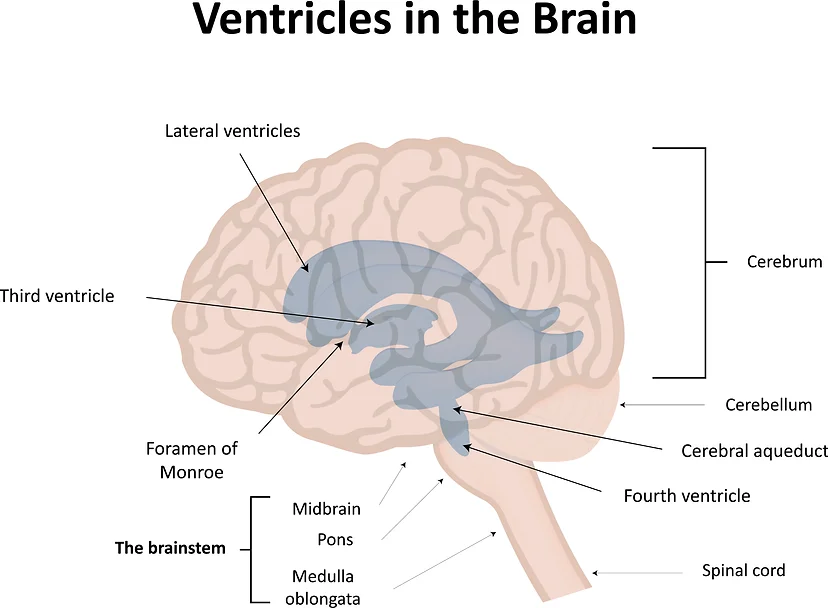
* Cornu anterius and Cornu posterius are found in the brain’s lateral ventricles. The anterior horn extends to the frontal lobe, the posterior horn to the occipital lobe. There is also an inferior horn extending to the temporal lobe.
